
- Neurologic Conditions
- ESWT
- Research
- 9 mins
Pain is a complex sensory and emotional experience that normally serves to protect the body from harm (Raja et al., 2020; Woolf, 2010). It is usually associated with nociception, the neural process of encoding potentially damaging stimuli (Basbaum et al., 2009), although pain and nociception are distinct processes. Chronic pain, however, can persist beyond its protective role and become a debilitating condition in its own right (Treede et al., 2019). One emerging non-invasive treatment for chronic musculoskeletal pain is extracorporeal shockwave therapy (ESWT). This post explores how nociception and pain perception work, and how ESWT influences these pathways.
Understanding Nociception and Pain
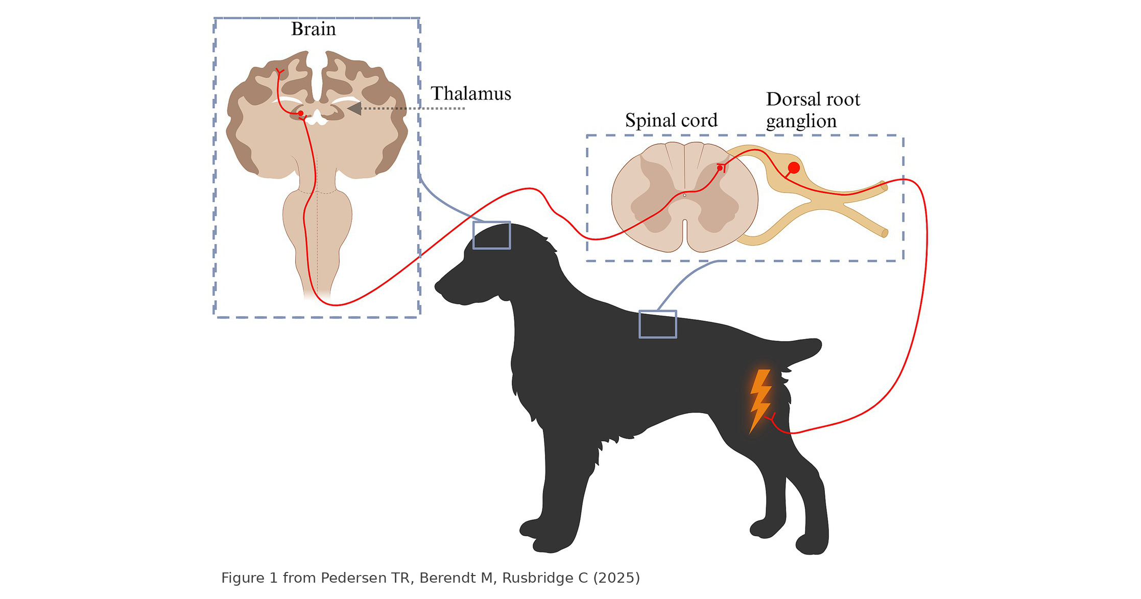
Nociception begins with specialized nerve endings called nociceptors, which detect noxious stimuli such as extreme temperatures, mechanical pressure, or chemical irritation. These nociceptors are present in skin, muscles, joints, and fascia. When activated, they send signals along primary afferent neurons to the dorsal horn of the spinal cord. From there, the signals ascend to higher brain centers, including the thalamus and somatosensory cortex, for pain perception (Pedersen et al., 2025).
Pain can be classified as acute or chronic. Acute pain is a short-term response to injury, whereas chronic pain persists due to sustained nociceptive input, central sensitization, or maladaptive neuroplastic changes (Parker, 2024). Chronic musculoskeletal pain, such as myofascial pain syndrome, often involves sensitization of nociceptors in the fascia, contributing to widespread pain and dysfunction (Ryskalin et al., 2022; Tesarz et al., 2011).
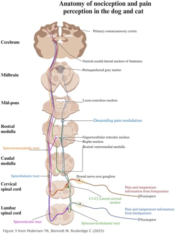 Nociception is not the same as pain. Nociceptive signals are the raw input, but the actual experience of pain is constructed in the brain (Raja et al., 2020). There, a distributed network of regions, including the somatosensory cortex, thalamus, limbic system, insula, and prefrontal cortex, interprets these signals and gives them meaning (Tracey & Mantyh, 2007). This network integrates sensory information with emotion, memory, attention, and prior experience (Bushnell et al., 2013; Wiech, 2016). Because the network is shaped by each patient’s genetics, previous injuries, learned associations, and even current emotional state, no two brains process the same nociceptive input in exactly the same way (Coghill et al., 2003; Mogil, 2012). Identical tissue damage can produce very different pain experiences from one patient to the next, and the same patient may perceive pain differently over time (Tracey & Mantyh, 2007).
How Shockwave Therapy Modulates Pain Perception
ESWT applies high-energy acoustic waves to targeted tissues, inducing mechanical and biological effects that alleviate pain. Several mechanisms are thought to explain its analgesic effects:
1. Fewer Nociceptive Nerve Fibers: Animal studies suggest that ESWT selectively reduces unmyelinated sensory nerve fibers in the treated area, which dampens pain transmission (Hausdorf, Lemmens, Heck, et al., 2008; Ryskalin et al., 2022).
2. Lower Levels of Pain-Related Neuropeptides: ESWT decreases substance P and calcitonin gene-related peptide (CGRP), key mediators of pain and neurogenic inflammation, in the dorsal root ganglion neurons that supply the treated area (Hausdorf, Lemmens, Kaplan, et al., 2008; Takahashi et al., 2003).
3. Enhanced Tissue Regeneration: By stimulating angiogenesis and cellular repair processes, ESWT contributes to the healing of damaged musculoskeletal structures, which may reduce the source of nociceptive input over time (D’Agostino et al., 2015; Wang et al., 2003).
4. Less Input Driving Central Sensitization: Chronic pain conditions involve heightened excitability of dorsal horn neurons, known as central sensitization (Woolf, 2010). By reducing nociceptive fibers and neuropeptide signaling in the periphery, ESWT may lessen the input that sustains this sensitization, although direct evidence of central effects is still limited (Ryskalin et al., 2022).
5. Improved Fascial Function: Fascia is densely innervated, including by nociceptive fibers, which makes it a plausible target in myofascial pain (Tesarz et al., 2011). ESWT may help by reducing fascial sensitivity and inflammation, but this effect has not yet been shown directly in fascial nociceptors (Ryskalin et al., 2022).
Clinical Applications and Future Directions
ESWT has shown promise in treating conditions such as plantar fasciitis, tendinopathies, and myofascial trigger points. Although the precise molecular mechanisms are still being investigated, its analgesic effects make ESWT a valuable tool in pain management. Future research may further clarify how ESWT influences neuroplasticity and nociceptive processing, potentially expanding its use beyond musculoskeletal disorders to neuropathic pain and central pain syndromes.
Because pain is ultimately a product of each individual’s nervous system, ESWT is most effective when tailored to the patient rather than applied as a one-size-fits-all protocol. Treatment parameters (energy level, number of pulses, treatment frequency, and anatomical targeting) should reflect the patient’s condition, pain presentation, tissue response, and stage of healing. Clinicians should reassess and adjust as the patient’s response evolves over the course of care.
Conclusion
Pain is a complex phenomenon rooted in nociceptive processing and central integration. ESWT offers a non-invasive option for chronic musculoskeletal pain, particularly when conventional therapies have not provided enough relief. By acting on peripheral pain mechanisms and possibly reducing the input that sustains central sensitization, ESWT can be a useful part of a pain management plan tailored to the individual patient.
References:
Basbaum, A. I., Bautista, D. M., Scherrer, G., & Julius, D. (2009). Cellular and molecular mechanisms of pain. Cell, 139(2), 267–284. https://doi.org/10.1016/j.cell.2009.09.028
Bushnell, M. C., Čeko, M., & Low, L. A. (2013). Cognitive and emotional control of pain and its disruption in chronic pain. Nature Reviews Neuroscience, 14(7), 502–511. https://doi.org/10.1038/nrn3516
Coghill, R. C., McHaffie, J. G., & Yen, Y.-F. (2003). Neural correlates of interindividual differences in the subjective experience of pain. Proceedings of the National Academy of Sciences, 100(14), 8538–8542. https://doi.org/10.1073/pnas.1430684100
D’Agostino, M. C., Craig, K., Tibalt, E., & Respizzi, S. (2015). Shock wave as biological therapeutic tool: From mechanical stimulation to recovery and healing, through mechanotransduction. International Journal of Surgery, 24(Pt B), 147–153. https://doi.org/10.1016/j.ijsu.2015.11.030
Hausdorf, J., Lemmens, M. A. M., Heck, K. D. W., Grolms, N., Korr, H., Kertschanska, S., Steinbusch, H. W. M., Schmitz, C., & Maier, M. (2008). Selective loss of unmyelinated nerve fibers after extracorporeal shockwave application to the musculoskeletal system. Neuroscience, 155(1), 138–144. https://doi.org/10.1016/j.neuroscience.2008.03.062
Hausdorf, J., Lemmens, M. A. M., Kaplan, S., Marangoz, C., Milz, S., Odaci, E., Korr, H., Schmitz, C., & Maier, M. (2008). Extracorporeal shockwave application to the distal femur of rabbits diminishes the number of neurons immunoreactive for substance P in dorsal root ganglia L5. Brain Research, 1207, 96–101. https://doi.org/10.1016/j.brainres.2008.02.013
Mogil, J. S. (2012). Pain genetics: Past, present and future. Trends in Genetics, 28(6), 258–266. https://doi.org/10.1016/j.tig.2012.02.004
Parker, R. L. (2024). Comparative analysis of chronic neuropathic pain and pain assessment in companion animals and humans. Frontiers in Veterinary Science, 11, 1520043. https://doi.org/10.3389/fvets.2024.1520043
Pedersen, T. R., Berendt, M., & Rusbridge, C. (2025). Neuroanatomy of spinal nociception and pain in dogs and cats: A practical review for the veterinary clinician. Frontiers in Veterinary Science, 12, 1534685. https://doi.org/10.3389/fvets.2025.1534685
Raja, S. N., Carr, D. B., Cohen, M., Finnerup, N. B., Flor, H., Gibson, S., Keefe, F. J., Mogil, J. S., Ringkamp, M., Sluka, K. A., Song, X.-J., Stevens, B., Sullivan, M. D., Tutelman, P. R., Ushida, T., & Vader, K. (2020). The revised International Association for the Study of Pain definition of pain: Concepts, challenges, and compromises. Pain, 161(9), 1976–1982. https://doi.org/10.1097/j.pain.0000000000001939
Ryskalin, L., Morucci, G., Natale, G., Soldani, P., & Gesi, M. (2022). Molecular mechanisms underlying the pain-relieving effects of extracorporeal shock wave therapy: A focus on fascia nociceptors. Life, 12(5), 743. https://doi.org/10.3390/life12050743
Takahashi, N., Wada, Y., Ohtori, S., Saisu, T., & Moriya, H. (2003). Application of shock waves to rat skin decreases calcitonin gene-related peptide immunoreactivity in dorsal root ganglion neurons. Autonomic Neuroscience, 107(2), 81–84. https://doi.org/10.1016/S1566-0702(03)00134-6
Tesarz, J., Hoheisel, U., Wiedenhöfer, B., & Mense, S. (2011). Sensory innervation of the thoracolumbar fascia in rats and humans. Neuroscience, 194, 302–308. https://doi.org/10.1016/j.neuroscience.2011.07.066
Tracey, I., & Mantyh, P. W. (2007). The cerebral signature for pain perception and its modulation. Neuron, 55(3), 377–391. https://doi.org/10.1016/j.neuron.2007.07.012
Treede, R.-D., Rief, W., Barke, A., Aziz, Q., Bennett, M. I., Benoliel, R., Cohen, M., Evers, S., Finnerup, N. B., First, M. B., Giamberardino, M. A., Kaasa, S., Korwisi, B., Kosek, E., Lavand'homme, P., Nicholas, M., Perrot, S., Scholz, J., Schug, S., . . . Wang, S.-J. (2019). Chronic pain as a symptom or a disease: The IASP Classification of Chronic Pain for the International Classification of Diseases (ICD-11). Pain, 160(1), 19–27. https://doi.org/10.1097/j.pain.0000000000001384
Wang, C.-J., Wang, F.-S., Yang, K. D., Weng, L.-H., Hsu, C.-C., Huang, C.-S., & Yang, L.-C. (2003). Shock wave therapy induces neovascularization at the tendon–bone junction. A study in rabbits. Journal of Orthopaedic Research, 21(6), 984–989. https://doi.org/10.1016/S0736-0266(03)00104-9
Wiech, K. (2016). Deconstructing the sensation of pain: The influence of cognitive processes on pain perception. Science, 354(6312), 584–587. https://doi.org/10.1126/science.aaf8934
Woolf, C. J. (2010). What is this thing called pain? Journal of Clinical Investigation, 120(11), 3742–3744. https://doi.org/10.1172/JCI45178