Evidence-Based Medicine for the PiezoWave2T Vet Shockwave
Evidence-based medicine is important to any medical field. It is the responsibility of the medical practitioners to read, evaluate, and interpret the available published literature. A few questions to consider are listed below.
- Are company-funded research studies of the same caliber as those that are done independent of corporate funding and input?
- What is the strength of the evidence being presented and could the interpretation be biased?
- The use of animal model systems for human healthcare is considered valid evidence-based medicine, can human research be applied to the veterinary patient?
- Is the cost/benefit ratio for funding clinical veterinary research appropriate for small companies?
 There is no right or wrong answer to any of the above questions. As with the treatment of veterinary rehabilitation patients, each situation is different. It is the responsibility of the veterinary practitioner to analyze and interpret the literature and make educated decisions for treatment plans based on the body of literature available.
This blog summarizes the different scientific evidence for the use of the PiezoWave2T Vet shockwave system and includes attachments to the specific publications.
The mechanism action for therapeutic shockwave is mechanotransduction and both mechanotransduction and the biological effects of mechanotransduction have been well studied and summarized in a previous blog. There is evidence medicine for the clinical use of PiezoWave shockwave therapy including two published papers using the PiezoWave2T Vet shockwave to treat canine shoulder injuries; case studies from Dr. Heather Owen, and a retrospective shoulder study using Dr. Julia Tomlinson’s. There are also publications that use PiezoWave shockwave for human musculoskeletal therapy. Some of the most pertinent ones include its use for treating: tennis elbow, calcific shoulder tendinopathy, and osteoarthritis of the knee.
Also, there is a recent comprehensive systematic review on shockwave use in animals in which they report both EFD values and bias scores from different publications. It is important to consider the dates of published literature as the understanding of shockwave treatment benefits progresses. In addition, changes have been made to the shockwave device technology. This evolution of shockwave technology optimizes the clinical applications, but it makes the results reported in earlier literature potentially difficult to duplicate. Finally, veterinary rehabilitation professionals should note the bias scores for some of the shockwave veterinary literature and be mindful of the conclusions that have been presented in papers with higher bias scores.
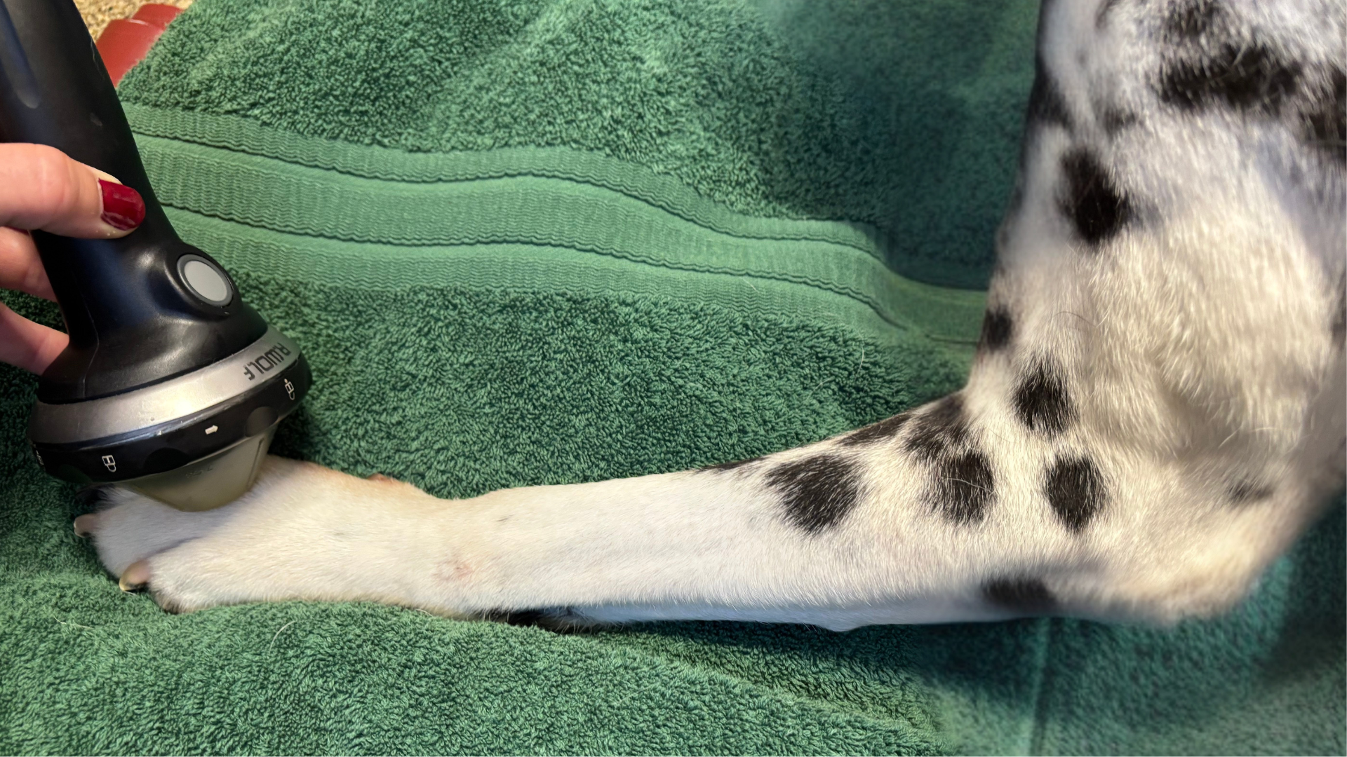
 An important fact about PiezoWave’s technology is that it is the only therapeutic modality that forms its active shockwave area within the tissue. The Richard Wolf therapy sources (trodes) for the PiezoWave2T Vet shockwave create an array of specifically aimed pressure waves that travel through the skin and innervated dermis and converge to form a shockwave within the tissue in a very controlled and consistent way. This is why there is no sensation as the PiezoWave’s energy passes through healthy tissue but is felt in injured tissue. This is also why the PiezoWave2T Vet shockwave provides such good biofeedback from animals. More details about the PiezoWave2T Vet and its therapy sources can be found in this blog.
With both electrohydraulic or electromagnetic shockwave systems, the shockwave itself is traveling from the base of the trode with a focal zone created by redirecting of the energy. These devices create a shockwave via either a spark plug or electromagnetic coil within the base of the trode. The released energy within the trode is then redirected to the focal zone via deflectors (Auersperg). In electrohydraulic and electromagnetic systems it is shockwaves that travel through the skin and other tissues until they reach the final focal zone. Thus, pain is felt as these shockwaves travel through the sensitive innervated dermis. For electrohydraulic shockwave, not only is the therapy painful as it passes through tissue to its final destination, but also the energy output from the physics of the spark gap method is inconsistent with some pulses having higher energy than others (Slezak et al). This inconsistency makes the therapeutic energy of electrohydraulic shockwaves difficult to quantify. Both inconsistent output, noise, and pain attribute to the need for some animals to be sedated when treated with these shockwave systems.
Are there any exact measurements available that verify the size and shape of the specific focal zone for the shockwave device you are currently using? ELvationUSA Vet believes the specificity of the focal zone is important. Treating the injury without involving healthy tissue/structures is an important part of the do-no-harm goal in medicine. Boasting that the treatment of a large area (including healthy tissue) simplifies the therapy application is contrary to the treatment goals and in reality, is a design limitation of the trode that is presented as a benefit even though it often results in post-treatment soreness.
There are many benefits to the PiezoWave shockwave systems including the large range of energy intensity (EFD) and depths of penetration that provide the ability to treat each animal in a specific manner as described in this blog and the unique specific therapy sources that often last for over 5 million pulses that are described in further detail here. However, for the best use of the PiezoWave 2T Vet shockwave, ElvationUSA Vet recommends that practitioners utilizing therapeutic shockwave have a solid understanding of the basic science as described within the ESWT guidelines from the ISMTS.
ELvation Marketing Team
Combining sales and flexible customer support with many years’ in-depth knowledge of medical equipment we offer customized solutions to create value with long-term investments and medical supplies. ELvation’s strength lies in its ability to combine the apparently contradictory needs of improving the standards of patient care by providing high-quality medical technology and good corporate profitability. We have a special partnership with Richard Wolf GmbH as their long-term authorized Sales & Service Team for piezo shockwave systems.


