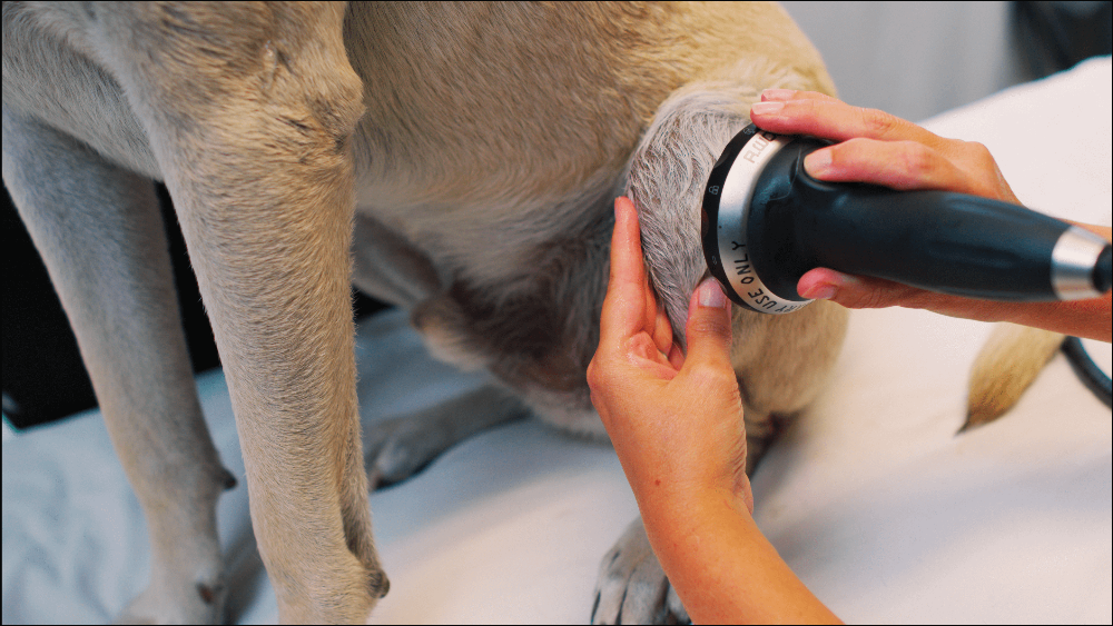
Optimizing PiezoWave2-Vet Therapy of The Canine Stifle
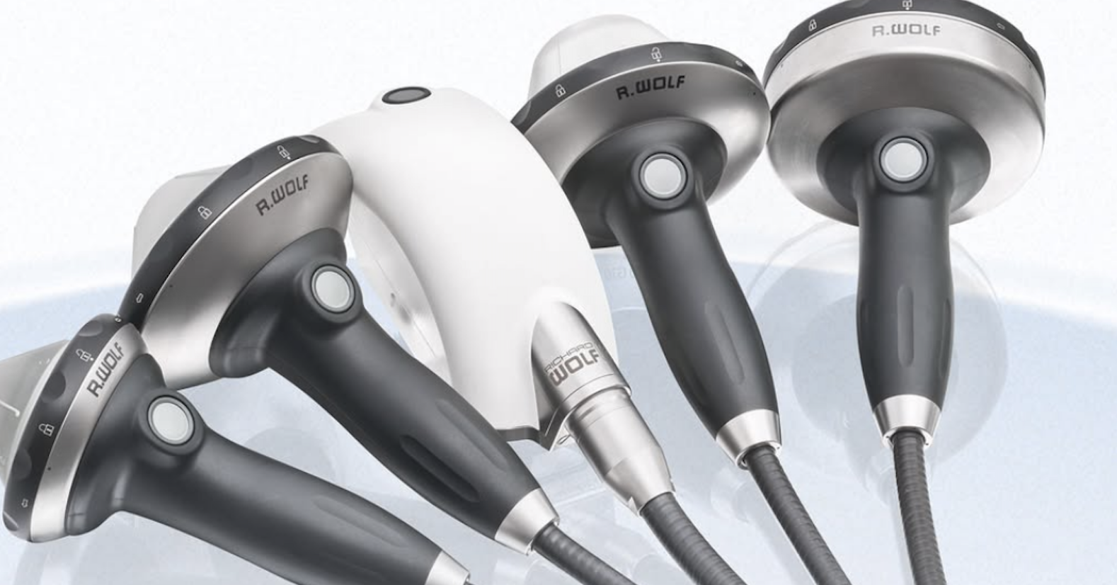
The PiezoWave2 provides specificity in the depth of penetration, energy output (EFD), and focal zone that allows the targeted tissue to be treated.
The canine stifle has many different structures that can be the source of pain. Both the structure needing treatment and the healing state of the tissue are taken into consideration when developing a treatment plan. It is also useful to assess surrounding muscles and other structures along the kinetic chain that can be affected due to acting as an antagonist on the injury or compensation from stifle pain.
Some of the specific structures of the stifle joint that can be treated with the PiezoWave2 are1 :
- synovium
- cranial cruciate ligament
- patellar ligament
- medial quadriceps tendon
- medial and lateral collateral ligaments
- long digital extensor tendon
- osteotomy site in a TPLO or TBLO
- subchondral bone for cartilage protection
It is important to understand the amount of energy required to initiate mechanotransduction is different for different tissue types. For example, bone is denser and requires a higher EFD than soft tissue structures. Acute injuries will require much less energy than chronic ones 2.
If a treatment plan involves multiple structures of the stifle, it is common to treat at different depths of penetration with different gel pads. Ultrasound imaging is the gold standard for depth determination; however, realistically most practitioners depend on excellent palpation skills and anatomical knowledge to determine tissue depth.
The effect of shockwave therapy on tendons and ligaments is well-documented 3. However, clinical studies on canine patients remain limited 4. Shockwave treatment of the stifle tendon and ligaments that are not within the joint capsule, including but not limited to the patellar ligament, and the medial and lateral collateral ligaments should be straightforward. The key to success on these soft tissue tendon and ligaments is the diagnosis and location of the injury. This is best done with diagnostic ultrasound imaging. The canine cranial cruciate ligament, CCL, is not only within the stifle joint capsule but also is positioned in cranial to caudal orientation making it difficult to position the ligament fibers specifically within the shockwave focal zone 1. It remains unknown if shockwave can actually regenerate the CCL when it has a partial tear. However, treatment should increase neovascularization, and aid in the healing process 5.
Shockwave therapy is known to have a chondroprotective effect through activation in the subchondral bone and there is literature that supports regeneration of cartilage with shockwave therapy 6. In canine patients, the PiezoWave depth and angle of entry can be targeted to the tibial subchondral bone to help in cartilage health. However, as previously mentioned activation of the bone via mechanotransduction requires higher energy (EFD) and can be uncomfortable. When choosing to target subchondral bone for cartilage treatment of the stifle, the practitioner should take the age and current condition of the stifle into account. In situations, where the joint is near to bone on bone and/or the dog is senior, treating for tissue changes in the cartilage may not be appropriate, whereas, in young and middle-aged dogs it may be helpful to their long-term mobility.
Shockwave therapy has been shown to improve lameness in dogs with osteoarthritis, OA, of the stifle 7. For AO of the stifle, simply treating the synovial fluid from one or two entry points along the joint line seems to be extremely beneficial. The molecular changes of the synovial fluid in arthritic joints are significant and shockwave has been shown to have a positive effect on the synovial fluid 8,9.
It has been shown that shockwave targeted to the osteotomy site post-TPLO surgery accelerates bone healing 10. When treating patients with implants, the specificity of the PiezoWave2 allows positioning of the focal zone such that it does not hit the implants. If the pressure waves do hit metal, they are reflected and the focal zone is disrupted. If the implant is secure in healthy bone, shockwave at intensities in the 0.15 mJ/mm2 energy range should not affect the implant, however, it may feel uncomfortable to the dog.
Optimization of PiezoWave2 protocols need to be case-specific and requires both knowledge of the anatomy and diagnosis of the injury. ELvationUSA has a suggested parameter table (https://hubs.ly/Q02kBk6r0) to help guide treatment plans, but we encourage the practitioners to develop the protocols that work best for their patients, technicians, and business plans.
References
1. Carpenter Jr, D. H., & Cooper, R. C. (2000). Mini review of canine stifle joint anatomy. Anatomia, histologia, embryologia, 29(6), 321-329. https://hubs.ly/Q02l04m402. “Is there an optimal dosage for veterinary shockwave?” Shockwave Resources Blog, ElvationUSA-Vet, June 21,2023. https://veterinaryshockwaveresources.com/blog/science-to-practice-the-optimal-dosage-for-veterinary-shockwave or http://bit.ly/3SBFFBC
3. Notarnicola, A., & Moretti, B. (2012). The biological effects of extracorporeal shock wave therapy (eswt) on tendon tissue. Muscles, ligaments and tendons journal, 2(1), 33. https://hubs.ly/Q02lk9Dj0
4. Alvarez, L. X., Repac, J. A., Kirby Shaw, K., & Compton, N. (2022). Systematic review of postoperative rehabilitation interventions after cranial cruciate ligament surgery in dogs. Veterinary Surgery, 51(2), 233-243. https://hubs.ly/Q02lk91-0
5. Wang, C. J., Wang, F. S., Yang, K. D., Weng, L. H., Hsu, C. C., Huang, C. S., & Yang, L. C. (2003). Shock wave therapy induces neovascularization at the tendon–bone junction. A study in rabbits. Journal of Orthopaedic Research, 21(6), 984-989. https://hubs.ly/Q02lkbD30
6. Chou, W. Y., Cheng, J. H., Wang, C. J., Hsu, S. L., Chen, J. H., & Huang, C. Y. (2019). Shockwave targeting on subchondral bone is more suitable than articular cartilage for knee osteoarthritis. International Journal of Medical Sciences, 16(1), 156 https://hubs.ly/Q02lkhw30
7. Dahlberg, J., Fitch, G., Evans, R. B., McClure, S. R., & Conzemius, M. (2005). The evaluation of extracorporeal shockwave therapy in naturally occurring osteoarthritis of the stifle joint in dogs. Veterinary and Comparative Orthopaedics and Traumatology, 18(03), 147-152. https://bit.ly/49GnIbZ
8. Watt, F. E., Hamid, B., Garriga, C., Judge, A., Hrusecka, R., Custers, R. J. H., ... & Vincent, T. L. (2020). The molecular profile of synovial fluid changes upon joint distraction and is associated with clinical response in knee osteoarthritis. Osteoarthritis and cartilage, 28(3), 324-333. https://hubs.ly/Q02l0LBd0
9. Frisbie, D. D., Kawcak, C. E., & McIlwraith, C. W. (2004). Evaluation of extracorporeal shock wave therapy for osteoarthritis. Evaluation, 1438, 1204. https://hubs.ly/Q02l0dw_0
10. Kieves, N. R., MacKay, C. S., Adducci, K., Rao, S., Goh, C., Palmer, R. H., & Duerr, F. M. (2015). High energy focused shock wave therapy accelerates bone healing. Veterinary and Comparative Orthopaedics and Traumatology, 28(06), 425-432. https://hubs.ly/Q02cJRWQ0
Video Courtesy of Dr. Debbie Gross Torraca, DPT, MSPT, CCRP, CCMT of Wizard of Paws.

ELvation Marketing Team
Combining sales and flexible customer support with many years’ in-depth knowledge of medical equipment we offer customized solutions to create value with long-term investments and medical supplies. ELvation’s strength lies in its ability to combine the apparently contradictory needs of improving the standards of patient care by providing high-quality medical technology and good corporate profitability. We have a special partnership with Richard Wolf GmbH as their long-term authorized Sales & Service Team for piezo shockwave systems.