
History
Extracorporeal shockwave therapy (ESWT) was first introduced for lithotripsy (kidney stone fragmentation) in the 1980s. Early observations of cortical bone remodeling in shockwave-treated areas led to its adoption in orthopedics1. Over the past three decades, ESWT has become an evidence-based treatment for delayed fracture healing, nonunions, and bone regeneration in both human and veterinary medicine2.
Mechanism of Action
Focused ESWT (f-ESWT) promotes bone healing through the process of mechanotransduction—the conversion of mechanical forces into biochemical signals3. In osteoblasts: integrins, ion channels, and cytoskeletal structures sense mechanical stress, triggering calcium influx and activation of MAPK/ERK signaling pathways. This cascade leads to the release of ATP, nitric oxide, and prostaglandins, along with cytoskeletal reorganization and activation of transcription factors such as RUNX2 and YAP/TAZ. In osteoblasts, these intracellular pathways ultimately enhance osteogenesis and mineralization.
Key molecular effects include:
- Activation of signaling pathways: FAK, ERK/MAPK, and mTOR pathways4
- Upregulation of osteogenic transcription factors: RUNX-2, CBFA15,6,7.
- Stimulation of angiogenesis: Upregulation of VEGF and HIF-1α expression8.
- Recruitment of mesenchymal stem cells: Enhancing differentiation into osteoblasts9,5.
Veterinary Applications
ESWT has proven effective in veterinary orthopedics for stimulating bone repair. Clinical and preclinical studies report accelerated bone healing in dogs, horses, and other species2. Key veterinary applications include:
- Post-surgical healing: Improved outcomes following TPLO procedures10.
- Fracture repair: Successful treatment of non-union fractures in animal models and clinical cases11,12.
- Enhanced bone metabolism: Support for osteogenesis via modulation of inflammation and angiogenesis13.
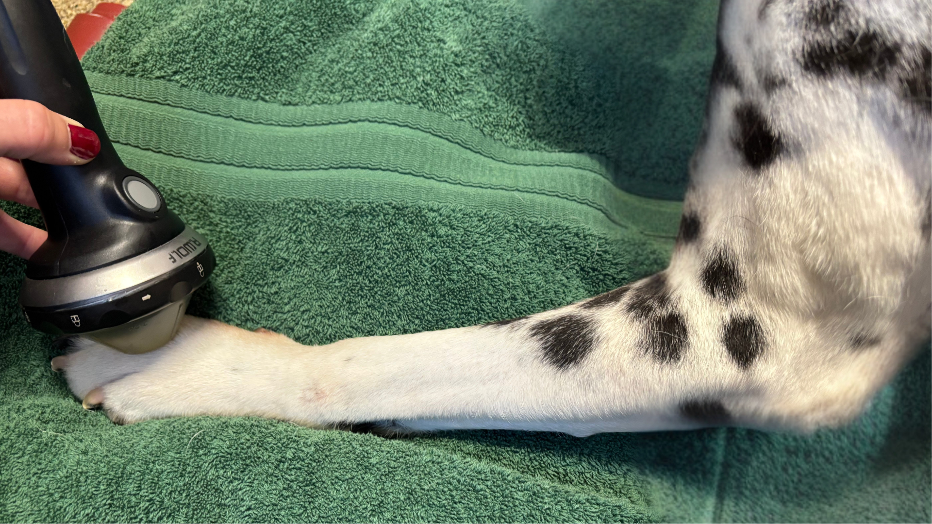
PiezoWave 2T Vet Shockwave Case Studies
 - Case 1 – Pomeranian with Radius/Ulna non-healing bone: Link to full case study
A 3.3 lb Pomeranian experienced 8 months of failed healing with only intermittent weight-bearing. After three PiezoWave treatments (1000 shocks, 0.191 mJ/mm²), callus formation was evident. After seven treatments, significant bone regeneration occurred, and within 3 months, the dog was fully weight-bearing with ongoing bone remodeling.
 - Case 2 – Australian Shepherd Digit Fracture: Link to full case study
An 18.6 kg Australian Shepherd presented with an unhealed digit fracture after 8 weeks. One PiezoWave session resolved lameness, and callus formation was seen after four weekly treatments. Notably, during these initial treatments, the energy flux density (EFD) remained below 0.063 mJ/mm². Most of the early literature reports a much higher EFD, so the effective lower EFD in this case study challenges previous experiences. Treatment was continued until near-complete bone healing was radiographically confirmed, allowing a successful return to agility competition.
These cases highlight the effectiveness of PiezoWave shockwave therapy in stimulating osteogenesis and achieving clinically significant outcomes with case-specific treatment parameters.
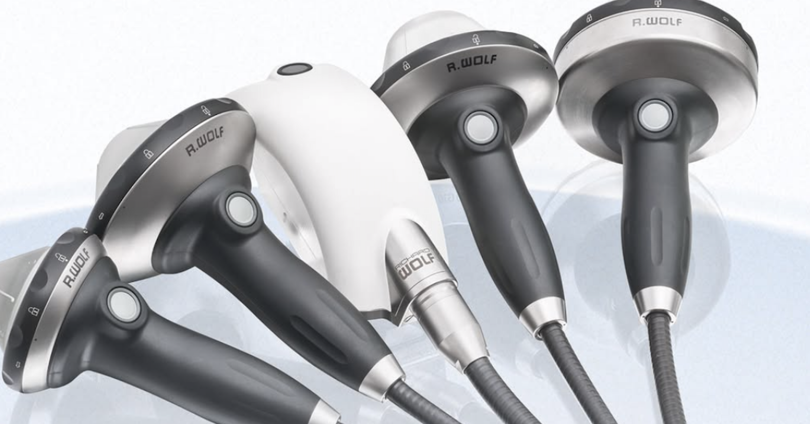
Benefits of PiezoWave Vet Over Other Systems
Piezoelectric technology provides distinct advantages:
- Precision: Sharply focused energy minimizes collateral tissue trauma.
- Patient comfort: Treatments are well tolerated without sedation, lowering risk and cost.
- Consistency: Piezoelectric crystals generate uniform energy, unlike electrohydraulic spark-gap devices.
- Cost-effectiveness: Long-lasting therapy sources (>5 million pulses) eliminate recurring trode replacement costs.
- Durability: Richard Wolf manufacturing ensures system longevity (10–12+ years).
Conclusion
Focused ESWT is a powerful, non-invasive therapy for bone healing in veterinary medicine. By stimulating key mechanotransduction pathways, it promotes callus formation, angiogenesis, and remodeling. PiezoWave Vet provides a precise, patient-friendly, and scientifically validated solution for managing challenging orthopedic cases.
References
1. Mittermayr, R., Haffner, N., Feichtinger, X., & Schaden, W. (2021). The role of shockwaves in the enhancement of bone repair. Injury, 52(S2), S84–S9.
2. Sansone, V., Ravier, D., Pascale, V., Applefield, R., Del Fabbro, M., & Martinelli, N. (2022). Extracorporeal shockwave therapy in the treatment of nonunion in long bones: A systematic review and meta-analysis. Journal of Clinical Medicine, 11, 1977.
3. Stewart, S., Darwood, A., Masouros, S., Higgins, C., & Ramasamy, A. (2020). Mechanotransduction in osteogenesis. Bone Joint Res, 9(1), 1–14.
4. Buarque de Gusmão, C. V., Batista, N. A., Vidotto Lemes, V. T., Maia Neto, W. L., de Faria, L. D., & Alves, J. M. (2019). Effect of low-intensity pulsed ultrasound stimulation, extracorporeal shockwaves and radial pressure waves on Akt, BMP-2, ERK-2, FAK and TGF-β1 during bone healing in rat tibial defects. Ultrasound in Medicine & Biology, 45(8), 2140–2161.
5. Hu, J., Liao, H., Ma, Z., Chen, H., Huang, Z., & Zhang, Y. (2016). Focal adhesion kinase signaling mediated the enhancement of osteogenesis of human mesenchymal stem cells induced by extracorporeal shockwave. Scientific Reports, 6, 20875.
6. Wang, F. S., Wang, C. J., Sheen-Chen, S. M., Kuo, Y. R., Chen, R. F., & Yang, K. D. (2002). Superoxide mediates shock wave induction of ERK-dependent osteogenic transcription factor (CBFA1) and mesenchymal cell differentiation toward osteoprogenitors. Journal of Biological Chemistry, 277(13), 10931-10937.
7. Hsu, C. C., Cheng, J. H., Wang, C. J., Ko, J. Y., Hsu, S. L., & Hsu, T. C. (2020). Shockwave therapy combined with autologous adipose-derived mesenchymal stem cells is better than with human umbilical cord Wharton’s jelly-derived mesenchymal stem cells on knee osteoarthritis. International Journal of Molecular Sciences, 21(4), 12177.
8. Tepeköylü, C., Wang, F. S., Kozaryn, R., Albrecht-Schgoer, K., Theurl, M., Schaden, W., & Holfeld, J. (2013). Shock wave treatment induces angiogenesis and mobilizes endogenous CD31/CD34-positive endothelial cells in a hindlimb ischemia model: implications for angiogenesis and vasculogenesis. The Journal of Thoracic and Cardiovascular Surgery, 146(4), 971-978.
9. Chen, Y. J., Wurtz, T., Wang, C. J., Kuo, Y. R., Yang, K. D., Huang, H. C., & Wang, F. S. (2004). Recruitment of mesenchymal stem cells and expression of TGF-β1 and VEGF in the early stage of shock wave–promoted bone regeneration of segmental defect in rats. Journal of Orthopaedic Research, 22(3), 526-534.
10. Kieves, N. R., MacKay, C. S., Adducci, K., Rao, S., Goh, C., Palmer, R. H., & Duerr, F. M. (2015). High energy focused shock wave therapy accelerates bone healing. Vet Comp Orthop Traumatol, 28, 425–432.
11. Johannes, E. J., Kaulesar, D. M., & Matura, E. (1994). High-energy shock waves for the treatment of nonunions: an experiment on dogs. Journal of Surgical Research, 57(2), 246-252.
12. Oginska, O. J., Whitelock, R., Hausler, K., Stelman, A., & Allen, M. J. (2019). Extracorporeal shock wave therapy for the treatment of non-union of a canine mandibular fracture. VCOT Open, 2(02), e56-e59.
13. Medina, C. (2023). Shockwave therapy in veterinary rehabilitation. The Veterinary Clinics of North America: Small Animal Practice, 53(4), 775–781.