
The equine sacroiliac (SI) joint is a complex, low-motion structure connecting the sacrum to the ilium within the pelvis. It plays a critical role in transferring propulsive forces from the hind limbs to the axial skeleton, particularly during high-performance activities such as galloping, jumping, and collection. Pain in this region is a common cause of poor performance in sport horses.
Function
The SI joint allows limited but essential multidirectional movement—rotation, shear, and sagittal tilting—for shock absorption and dynamic coordination. Key functions include:
- Stability via interlocking articular surfaces, robust interosseous ligaments, and strong muscular support.
- Force transmission from hind limbs through the pelvis to the spine.
- Shock absorption during limb-ground interaction.
- Locomotor coordination with the lumbar spine for symmetrical gait
Structure
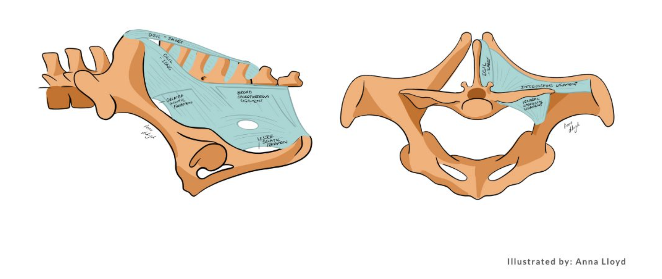
An amphiarthrodial-synovial joint, the SI joint consists of paired synovial compartments between the articular surfaces of the sacrum and ilium. These are surrounded by fibrous structures and reinforced by:
- Dorsal sacroiliac ligament (sacrum to ilium/sacral tuberosity)
- Ventral sacroiliac ligament (ventral joint capsule)
- Interosseous ligament (major stabilizer, limiting motion)
Surrounding musculature to the SI joint region includes longissimus dorsi, gluteals, iliopsoas, and hamstrings. These muscles further stabilize and indirectly influence the movement and function of the SI joint.
Injections
SI region injections are used diagnostically to localize pain and therapeutically to reduce inflammation. Signs of SI-related pain include asymmetrical pelvic musculature, reluctance to collect, and gait abnormalities.
Direct intra-articular injections are rarely performed due to the deep anatomical location and irregular joint surfaces. Instead, ultrasound-guided periarticular injections are standard, typically using long spinal needles and injecting corticosteroids (e.g., triamcinolone) and local anesthetics (e.g., mepivacaine).
Shockwave Therapy
Methods for treating the SI joint all encounter access challenges. An ultrasound-guided needle can get close to the synovial joint but the process is not an inter-articular joint injection. It requires both targeted angles of entry as well as passing the needle by specific boney landmarks and through various different muscles and ligaments.
Although shockwave therapy is effective in managing SI region pain and inflammation, a shockwave’s focal zone is unlikely to reach the true synovial joint due to anatomical barriers. When the shockwaves encounter tissues that all have differing acoustic impedances (bones, ligaments, and different muscles), the shockwave energy will be attenuated by these tissues and may not reach the location of the periarticular injections or the synovial joint. Because of these limitations, the goal for shockwave treatments for the SI joint are related to the treatment of the soft tissue surrounding the joint rather than the joint itself.
Shockwave therapy to the SI joint region is highly effective. When using the PiezoWave2T Vet system combined with practitioner-guided scanning and equine (patient) biofeedback, shockwaves effectively target reactive soft tissues in the sacral region. When shockwaves are applied using this method they help improve function and reduce pain, even without directly treating the synovial compartment and surrounding fibrous structures. They accomplish this by reducing muscle spasms due to guarding and improving blood flow to restricted area within soft tissue, as well as provide anti-inflammatory chemicals through mechanotransduction.
Summary
Shockwave therapy in the sacroiliac region is clinically valuable for managing equine back and pelvic pain. While the actual SI joint is rarely the direct target, strategic treatment of surrounding tissues—guided by clinical assessment and equine response—yields significant functional improvement. Learn more about PiezoWave: Small animal providers click here, and large animal providers click here.
References
- d'Agostino, M. C., Craig, K., Tibalt, E., & Respizzi, S. (2015). Shock wave as biological therapeutic tool: From mechanical stimulation to recovery and healing, through mechanotransduction. International journal of surgery, 24, 147-153.
- Denoix, J. M. (1999). Functional anatomy of the equine pelvic limb. *Veterinary Clinics of North America: Equine Practice*, 15(2), 283–306. https://doi.org/10.1016/S0749-0739(17)30101-1.
- Denoix, J. M., & Dyson, S. J. (2013). Diagnosis and management of lameness in the horse (2nd ed.). Elsevier Saunders.
- Dyson, S., Tranquille, C., Collins, S., & Murray, R. (2011). The sacroiliac joints: comparative evaluation of imaging methods. *Equine Veterinary Education*, 23(9), 489–498. https://doi.org/10.1111/j.2042-3292.2010.00266.
- Goff, L., Jeffcott, L. B., & Barneveld, A. (2008). Comparative biomechanics and histology of the sacroiliac joint in horses. *Equine Veterinary Journal*, 40(2), 166–172. https://doi.org/10.2746/042516408X245192.
- Haussler, K. K. (1999). Diagnosis and treatment of sacroiliac dysfunction. Veterinary Clinics of North America: Equine Practice, 15(1), 199–219. https://doi.org/10.1016/S0749-0739(17)30197-4.